Ingredients
- What is Pectus Carinatum? Why Does It Happen?
- Pigeon Chest Formation Mechanism and Incidence
- Types of Pectus Carinatum: Chondrogladios and Chondromanubrial
- Pigeon Chest Symptoms and Physical Effects
- Protrusion and Deformity of the Chest Wall
- Exercise Intolerance and Shortness of Breath
- Pigeon Chest Diagnosis and Evaluation Methods
- Physical Examination and State Index
- Radiological Imaging: Thorax CT and MRI
- Pectus Carinatum Treatment Options
- Non-Surgical Treatment: Use of Orthotics (Corset) and Pressure Adjustment
- Success Rates and Duration of Use in Orthotic Treatment
- Pectus Carinatum Surgery (Surgical Methods)
- Closed Surgery (Abramson Technique): Minimally Invasive Approach
- Open Surgery (Modified Ravitch Technique): Who is it applied to?
- Post-Treatment Recovery Process and Quality of Life
- Psychological Effects and Self-Confidence Gain
- Frequently Asked Questions
- Does Pigeon Chest Go Away On Its Own?
- What is the Best Age Range for Orthotic Treatment?
- When is the bar removed after Abramson’s surgery?
- Scientific Bibliography
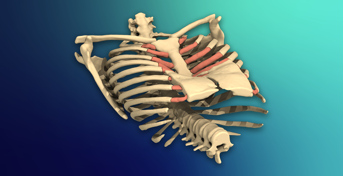
Pectus carinatum is a deformity characterized by the outward growth of the sternum bone (sternum) that forms the anterior chest wall and the cartilage ribs connected to it.
This condition, popularly known as “pigeon chest”, manifests itself when the rib cage tapers forward, as if it were the chest of a bird.
It is the second most common structural disorder among chest wall deformities after shoemaker’s chest (pectus excavatum).
While it often goes unnoticed at birth, it can become apparent during the rapid growth spurt of adolescence, leading to both physical and psychological issues.
Today, thanks to the developing technology, the treatment of this condition is successfully carried out with special devices called “orthoses” or closed surgery techniques without the need for surgery.
What is Pectus Carinatum? Why Does It Happen?
Pectus carinatum occurs when the sternum pushes forward as a result of abnormal and uncontrolled elongation of the rib cartilages.
Although the exact cause of this structural disorder is not known exactly, metabolic processes or growth factors in cartilage tissue are thought to be effective.
A family history of a similar rib cage disorder is found in about 25% of cases, indicating a strong genetic predisposition as a strong factor.
In addition, its incidence is quite high with other skeletal system disorders such as scoliosis (curvature of the spine).
Pigeon Chest Formation Mechanism and Incidence
The mechanism of its formation is based on the irregularity in the ossification process of cartilage tissue and the fact that this tissue acts as a lever that lifts the sternum outwards.
According to statistical data, it is approximately 4 times more common in boys than girls.
It usually becomes evident between the ages of 11-15, when body development is at its fastest, to be seen from the outside with the naked eye.
Types of Pectus Carinatum: Chondrogladios and Chondromanubrial
Medically, pigeon chest is divided into two main groups according to the location of the protrusion:
- Chondrogladiolar Type: It is the most common type; The middle and lower parts of the sternum taper outward.
- Chondromanubrial Type: It is a rarer type and more complex to treat; It occurs when the upper part of the breastbone (manubrium) protrudes and is also called the “pigeon fly”.
In both types, the deformity can be symmetrical, as well as asymmetrical forms where only one side of the chest protrudes.
Pigeon Chest Symptoms and Physical Effects
Although the main problem in most cases is aesthetic appearance, the loss of flexibility of the rib cage can lead to physical limitations.
Protrusion and Deformity of the Chest Wall
The first and most obvious symptom is a hard, bony protrusion that forms on the front of the rib cage.
This protrusion can be visible even under tight clothing and can trigger behavioral disorders in children, such as avoidance of social environments and tendency to slouch over.
Exercise Intolerance and Shortness of Breath
Since the rib cage is fixed in the “continuously inhaled” position in the pectus carinatum, the stretching capacity of the lungs may be restricted.
Especially in young people who do active sports, fatigue, shortness of breath during exertion and sometimes stinging pain in the chest area can be seen.
Although pressure on the heart is rare, it can be difficult for the lungs to ventilate at full capacity due to the rigid structure of the rib cage.
Pigeon Chest Diagnosis and Evaluation Methods
The diagnostic process includes a physical examination, as well as radiological tests that measure the severity of the deformity.
Physical Examination and State Index
The specialist physician examines the patient’s rib cage from different angles and checks the symmetry and flexibility of the deformity.
The Haller Index is the ratio of the transverse diameter of the rib cage to its longitudinal diameter; This index numerically reveals the severity of the stenosis or protrusion.
Radiological Imaging: Thorax CT and MRI
Thorax CT (Computed Tomography) guides the treatment plan by mapping the cartilage and bone structures in detail.
Thanks to CT scans, the rotations (torsion) on the sternum and the position of the internal organs are clearly evaluated.
MRI (MRI) imaging may also be preferred to examine non-bone tissues in adolescents due to less radiation risk.
Pectus Carinatum Treatment Options
The most important revolutionary development in the treatment of pigeon chest is the prominence of non-surgical solutions at the age when the rib cage is flexible.
Non-Surgical Treatment: Use of Orthotics (Corset) and Pressure Adjustment
Orthotic treatment is a special corset system that is placed over the rib cage and applies controlled pressure from the outside.
Just like in braces treatment, it puts constant pressure on the bone and cartilage tissues, pushing the deformity inward.
Modern orthoses with pressure adjustment increase the patient’s comfort and allow the physician to follow the treatment process millimetrically.
Success Rates and Duration of Use in Orthotic Treatment
The success rate of orthotic treatment in young people with flexible chest walls is between 80% and 90%.
- Adaptation Process: In the first weeks, low-pressure applications are made to get the body used to the device.
- Usage Time: These devices, which should be worn for an average of 12-23 hours a day, are used between 6 months and 2 years, depending on the severity of the deformity.
- Tracking: With regular checks, the pressure points are updated and the amount of improvement is monitored.
Prof. Dr. Levent Alpay: “Age” is the most critical factor in the treatment of pectus carinatum. Orthotic treatment, which is started during adolescence when the rib cage is still flexible, can save the patient from a major surgery. However, when applied after ossification is completed, surgery may become the only option. For this reason, parents should consult a specialist as soon as they notice changes in their child’s breast structure, which directly affects the success of treatment.
Pectus Carinatum Surgery (Surgical Methods)
Surgical options are considered in patients who do not respond to orthotic treatment or whose bone structure is too hardened to be corrected with orthotics.
Modern medicine offers both closed and open techniques for reshaping the rib cage.
In the decision for surgery, the age of the patient, the type of deformity and the psychological state of the person are considered as a whole.
Closed Surgery (Abramson Technique): Minimally Invasive Approach
The Abramson technique is the version of the Nuss surgery for the shoemaker’s chest, developed for the pigeon’s chest.
A metal bar is inserted over the breastbone (sternum) through small incisions made on both sides of the chest.
This bar presses the outward protrusion inward, bringing the rib cage to its normal anatomical position.
Since it does not require large incisions, the aesthetic results are excellent and the hospital stay is quite short.
Open Surgery (Modified Ravitch Technique): Who is it applied to?
The Ravitch method is preferred in cases where the chest wall is too hard or the deformity is asymmetrical and excessively advanced.
In this procedure, abnormal cartilage ribs attached to the sternum are removed and the breastbone is brought to the plane where it should be.
It is generally the method that gives the most reliable results in adults who have completed their growth and development or in patients with complex bone structure.
Post-Treatment Recovery Process and Quality of Life
Whether the treatment is done with orthotics or surgery, the ultimate goal is the full recovery of the patient, both physically and socially.
After surgery, patients are usually discharged within a few days and can return to normal activities within a month.
The bars placed with closed surgery are removed with a simple procedure after the rib cage adapts to its new shape (average 2-3 years).
Psychological Effects and Self-Confidence Gain
Young people with pigeon chests often exhibit behaviors such as avoiding swimming, wearing loose clothing, and slouching due to their body image.
The improvement of the rib cage after treatment provides a rapid increase in self-confidence and more active participation in social life in these young people.
Physical recovery not only increases lung capacity but also has lasting positive effects on a person’s mental well-being.
Treatment Methods Comparison Table
| Feature | Orthosis (Corset) Treatment | Abramson Technique (Closed) | Ravitch Technique (On) |
| Method | Non-Surgical / Pressurized | Minimally Invasive (Metal Bar) | Open Surgery (Cartilage Removal) |
| Hospitalization | Not required | 2 – 4 Days | 4 – 6 Days |
| Age Range | 10 – 16 Years (Flexible period) | Adolescence and Young Adulthood | All ages (Especially advanced age) |
| Success Rate | 85%+ (When compliant) | %95+ | %90+ |
Prof. Dr. Levent Alpay: Success in the treatment of pigeon chest begins with the patient’s belief in the treatment. While the support of the family and the regular use of the device by the young person constitute 100% of the success in orthotic treatment, a team experienced in surgical options provides the best aesthetic appearance with the least risk. It should not be forgotten that this is not just an aesthetic concern, but the construction of a young person’s self-confidence and posture in the future.
Case Experience (Anonymous):
Our 14-year-old male patient had started to develop social phobia due to prominent pectus carinatum. Measurements showed that the chest wall was still flexible, and a personalized pressure-adjustable orthotic treatment was started instead of surgery. At the end of 1 year of regular use, our patient, whose rib cage returned to normal completely, regained his health and self-confidence without requiring any surgical intervention.
If you are concerned about changes in your child’s breast structure or if you want to know whether the current condition requires surgery, you can make an appointment with our clinic and seek expert opinion.
Frequently Asked Questions
Does Pigeon Chest Go Away On Its Own?
No, pectus carinatum is a developmental disorder and unless treated, the bone structure hardens in this way; however, it can continue to change shape until growth is complete.
What is the Best Age Range for Orthotic Treatment?
The best results are obtained between the ages of 10-15 when the rib cage is still flexible; However, in appropriate cases, success can be achieved up to the age of 18.
When is the bar removed after Abramson’s surgery?
It is generally recommended that the metal bar remain in the body for 2 to 3 years in order for the rib cage to ossify enough to maintain its new form.
Scientific Bibliography
- Journal of Pediatric Surgery: Compressive Orthotic Bracing for Pectus Carinatum
- The Annals of Thoracic Surgery: The Abramson Procedure: Minimally Invasive Repair of Pectus Carinatum
- PubMed (NCBI): Management of Pectus Deformities in Adolescents
- Pectus Clinic International: Non-surgical Treatment Protocols