Ingredients
- What is Chest Wall Surgery? Which Diseases Does It Cover?
- Chest Wall Tumors and Surgical Treatment
- Chest Wall Deformities (Pectus Excavatum and Carinatum)
- Chest Wall Trauma and Rib Fracture Repair
- Diaphragm Surgery and Functional Disorders
- The Relationship Between Diaphragm Paralysis and Shortness of Breath
- Diaphragm Plication Surgery (Stretching Repair)
- Diaphragmatic Hernia and Treatment Methods
- Diagnosis and Diagnostic Process: Radiological Evaluation
- Modern Techniques in Chest Wall and Diaphragm Surgeries
- Closed Methods (VATS and Robotic Diaphragm Surgery)
- Chest Wall Reconstruction and Patch Applications
- Postoperative Recovery and Rehabilitation Process
- Frequently Asked Questions
- Does Shortness of Breath Go Away Immediately After Diaphragm Surgery?
- Do Chest Wall Tumors Recur After Surgery?
- What are the Advantages of Closed Diaphragm Surgery?
- Scientific Bibliography
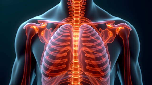
Chest wall and diaphragm surgery is a specialized specialty that aims to restore the structural integrity of the rib cage, which protects vital organs, and the functions of the diaphragm, the most important muscle of breathing.
While the rib cage protects the heart and lungs from external influences; The diaphragm muscle works like a pump under these organs, allowing us to breathe in and out.
Tumors, deformities, or loss of function in these areas can lead not only to aesthetic concerns but also to severe respiratory and circulatory failures.
Modern thoracic surgery approaches aim to both repair these complex anatomical structures and maximize their function.
Thanks to the advanced surgical techniques applied today, patients recover much faster and regain their respiratory capacity in a short time.
What is Chest Wall Surgery? Which Diseases Does It Cover?
Chest wall surgery; It is the surgical treatment of the area that includes the ribs, sternum and the muscle and soft tissues covering these bones.
This surgical field encompasses a wide range of diseases, from congenital disorders to aggressive tumors that develop later.
The main goal is to ensure that the rib cage continues to act as a protective shield and to create a space where the lungs can expand comfortably.
Chest Wall Tumors and Surgical Treatment
Chest wall tumors can originate directly from the ribs or sternum, or they can spread to this area from surrounding organs such as lung or breast cancer.
The basic principle in the surgical treatment of tumors is the complete removal of the mass along with the surrounding healthy tissue.
After the mass is removed, the cavity formed is reconstructed with the help of the patient’s own tissues or biosynthetic patches, preserving the integrity of the chest.
Chest Wall Deformities (Pectus Excavatum and Carinatum)
Deformities, popularly known as “shoemaker’s chest” (Pectus Excavatum) and “pigeon chest” (Pectus Carinatum), are among the most common areas of chest wall surgery.
These conditions not only lead to psychological problems; In advanced levels, it can put pressure on the heart, causing arrhythmias or fatigue.
In modern treatment, aesthetic and functional improvement is achieved thanks to metal bars placed in the rib cage with minimally invasive (closed) methods such as Nuss and Abramson.
Chest Wall Trauma and Rib Fracture Repair
Multiple rib fractures that occur in situations such as falling from a height or traffic accidents can lead to life-threatening conditions called “flail chest”.
In these patients, who used to be recommended only bed rest, today the process of fixing the ribs (osteosynthesis) is performed using special titanium plates and screws.
This procedure quickly relieves the patient’s pain, shortens the length of stay in the intensive care unit and minimizes the risk of lung collapse.
Diaphragm Surgery and Functional Disorders
The diaphragm is a thin but very strong layer of muscle that separates the chest cavity from the abdominal cavity.
Sagging, paralysis or holes in this muscle, which undertakes 70-80% of the breathing process alone, directly affect the quality of life.
The Relationship Between Diaphragm Paralysis and Shortness of Breath
As a result of damage to the “phrenic nerve” that commands the diaphragm muscle, the diaphragm sags upwards and loses its function.
This condition causes the abdominal organs (liver, stomach) below the diaphragm to go up and compress the lung.
Patients feel severe shortness of breath, especially when they lie on their backs or bend over.
Diaphragm Plication Surgery (Stretching Repair)
The treatment of the paralyzed diaphragm that hangs up like a balloon is a stretching surgery called “plication”.
The diaphragm muscle, which is loosened during the surgery, is folded with special sutures, stretched and lowered.
Thus, the area where the lung can expand is opened again and the patient’s breathing capacity increases instantly.
Diaphragmatic Hernia and Treatment Methods
Diaphragmatic hernias occur when organs in the abdomen pass through a hole in the diaphragm into the chest cavity.
These holes can be congenital (Bochdalek or Morgagni hernias), or they can develop after penetrating or blunt trauma.
The treatment is to push the herniated organs back into the abdomen and close the hole in the diaphragm with intact sutures or patches.
Diagnosis and Diagnostic Process: Radiological Evaluation
Advanced imaging techniques are as critical as physical examination in diagnosing chest wall and diaphragm problems.
- Dynamic Fluoroscopy (Sniff Test): It is used to monitor the movements of the diaphragm during breathing live.
- Computed Tomography (CT): It clarifies fractures in the bone structure, the depth of tumors and the size of hernias.
- MRI (Magnetic Resonance): It is used in the evaluation of soft tissue tumors and nerves.
- Pulmonary Function Tests (PFT): It measures how much diaphragm paralysis restricts lung capacity.
Modern Techniques in Chest Wall and Diaphragm Surgeries
Advances in medical technology have allowed these challenging surgical interventions to be performed with much smaller incisions and less damage.
Closed Methods (VATS and Robotic Diaphragm Surgery)
Diaphragm surgeries, which used to require the rib cage to be opened widely, can now be performed through 3 small holes (VATS) or with the help of robotic arms.
Robotic surgery allows even the deepest points of the diaphragm to be reached with a high-resolution image and the stitches to be placed much more firmly.
Chest Wall Reconstruction and Patch Applications
Highly engineered materials are used to close the cavities formed in the rib cage after major tumor surgeries.
Using titanium bars, synthetic mesh systems or the patient’s own muscle tissues (flaps), the chest wall is rebuilt in a way that is both strong and flexible.
Surgical Method Comparison Table
| Feature | Closed (VATS/Robotic) Surgery | Open Surgical Approach |
| Incision Size | Small holes of 1-2 cm | Large incision of 15-20 cm |
| Pain Level | Minimum / Low | Medium / High |
| Hospital Stay | 2 – 3 Days | 5 – 7 Days |
| Return to Daily Life | 1 – 2 Weeks | 4 – 6 Weeks |
Postoperative Recovery and Rehabilitation Process
The most important factor in the postoperative period is to ensure that the lungs inflate to full capacity.
Patients are started breathing exercises and walking programs the day after the surgery.
Patients who undergo surgery with the closed method can usually get rid of their drains within a few days and return home.
Prof. Dr. Levent Alpay: Many patients who suffer from shortness of breath due to diaphragmatic paralysis ignore the surgical solution, thinking that this condition is a “sign of aging” or “heart disease”. However, with a correct plication surgery, it is possible to change the patient’s quality of life in one day. The important thing is to analyze the degree of functional loss well and intervene with the least traumatic method.
Case Experience (Anonymous):
Left diaphragmatic paralysis was detected in a 62-year-old male patient who experienced shortness of breath while climbing stairs. After the diaphragm plication performed with the robotic method, the patient’s diaphragm was lowered to its normal anatomical level. The patient, who described an 80% improvement in shortness of breath the day after the surgery, was discharged on the 2nd day.
For your complaints of chest wall deformity, mass or shortness of breath caused by the diaphragm, you can make an appointment with our clinic for a detailed evaluation and modern surgical solutions and seek expert opinion.
Frequently Asked Questions
Does Shortness of Breath Go Away Immediately After Diaphragm Surgery?
Yes, especially after diaphragm plication surgery, patients immediately feel that breathing becomes easier as the pressure on the lung is removed.
Do Chest Wall Tumors Recur After Surgery?
It depends on the type of tumor and the cleanliness of the surgical margins; The risk of recurrence is very low in benign tumors removed with clean surgical margins.
What are the Advantages of Closed Diaphragm Surgery?
Less pain, shorter hospital stay, low risk of infection and small aesthetic scarring are the biggest advantages.
Scientific Bibliography
- The Annals of Thoracic Surgery: Chest Wall Reconstruction: Evolution and Current Concepts
- Journal of Thoracic Disease: Diaphragmatic Plication: Robotic and VATS Approaches
- PubMed (NCBI): Management of primary chest wall tumors
- Journal of Visceral Surgery: Diaphragmatic Hernia: Diagnosis and Surgical Management