
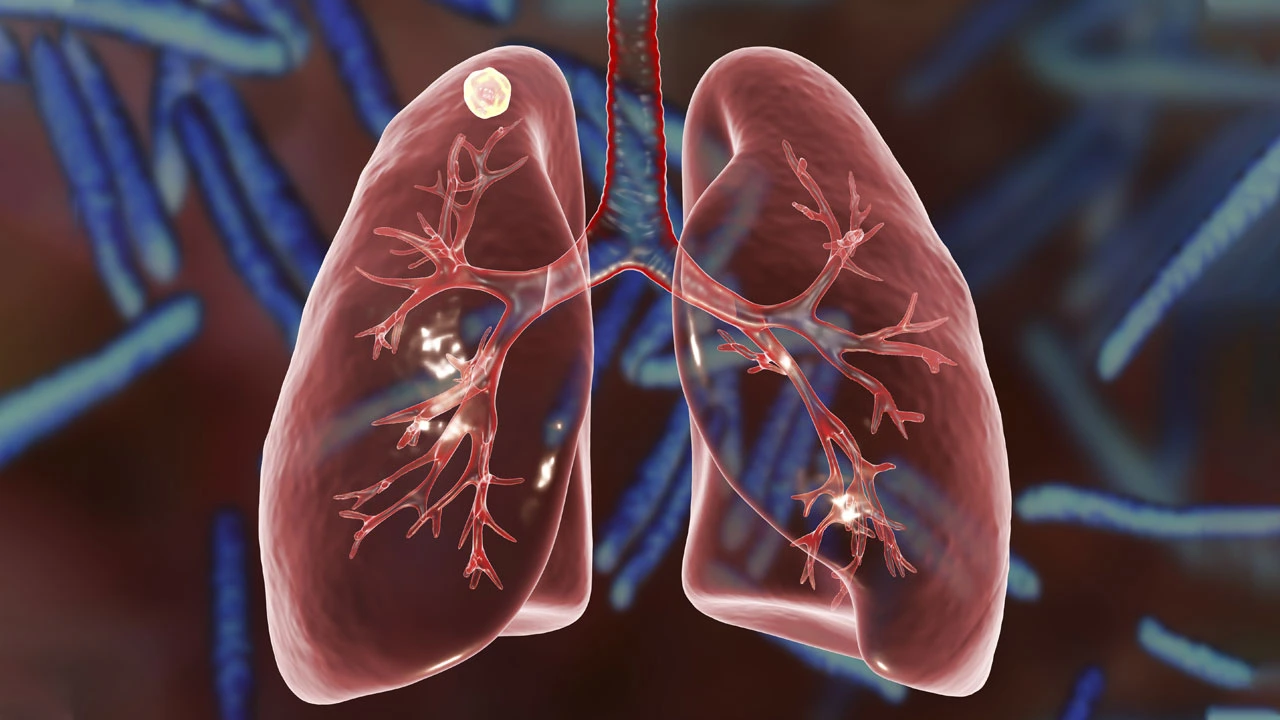
Lung nodules are detected much more frequently by chance today, thanks to developing radiological imaging techniques. Although the expression “lung nodule” seen in radiology reports causes serious concern among patients, the majority of these formations are benign. However, there are certain criteria that determine the potential of a nodule to be malignant. These criteria are; The radiological characteristics of the nodule are evaluated based on the patient’s personal health history and the rate of change over time.
Basic Criteria Determining the Risk of Cancer in Lung Nodules
To understand whether a lung nodule indicates cancer, physicians first analyze the morphological structure of the nodule. Although a feature alone does not make a diagnosis, the combination of certain features increases the risk scoring.
The Importance of Nodule Size and Millimetric Measurements
The diameter of the nodule is directly related to the risk of cancer. In the general medical literature, the risk of cancer in nodules under 5 mm is less than 1%, while this risk can exceed 60% in formations over 20 mm (2 cm). In particular, formations exceeding 30 mm (3 cm) are no longer called nodules but “masses” and are considered malignant until proven otherwise and require further examination.
Edge Structure: Is It Uniformly Bounded or Spiculate?
Benign nodules usually have sharp and smooth borders. On the other hand, if there are radial extensions called “spicular” or an irregular, serrated structure on the edges of the nodule, this may suggest that the nodule is trying to infiltrate the surrounding tissue. The “lobulated” (gnarled) margin structure is also among the radiological findings that increase the risk of cancer.
Density of the Nodule: Solid and Ground Glass (Subsolid) Appearance
Nodules are divided into three groups according to their radiological density:
- Solid Nodules: They contain completely dense tissue.
- Ground Glass Nodules: They are translucent views in which the vascular and bronchial structures under the lung tissue can be distinguished.
- Semi-Solid Nodules: They are structures with both frosted glass and dense solid components. Statistically, semi-solid nodules constitute the highest risk group for early-stage adenocarcinoma, especially if the solid component increases over time.
Critical Changes Indicating Cancer in the Follow-up Process of the Nodule
The behavior of a first detected nodule becomes clear during the follow-up process. Lung cancer is a dynamic process, and it’s vital to monitor change.
Growth Rate and Volume Doubling Time (VDT)
Cancer cells multiply at a certain rate. In lung cancers, the doubling time of the nodule’s volume is usually between 20 days and 400 days. If a nodule doubles in size in less than 15 days, it is usually a focus of infection (inflammation). If its size does not change at all for 2 years, it is most likely benign.
Comparative Analysis with Old Tomography Records
The most valuable procedure that can be done for a newly detected nodule is to access the patient’s tomography or x-ray records from 5-10 years ago, if any. A formation that has been the same size since years ago is extremely unlikely to be cancerous.
Evaluation of Contrast Uptake and PET-CT Results
PET-CT may be preferred to measure metabolic activity in nodules larger than 8 mm. However, PET-CT does not always give definitive results. Tuberculosis or fungal infections may also show high involvement (SUVmax value), and some types of cancer with a slow course may give false negative results.
Prof. Dr. Levent Alpay: Do not panic just by looking at the size of a nodule detected in your lung. The ‘character’ of the nodule is much more important than its size. Especially in ground-glass-looking nodules, the follow-up process can take years; For this reason, it is critical that you stick to the control schedule determined by your doctor.
Patient-Related High Risk Factors and Clinical Manifestations
Radiological findings should be combined with the clinical profile of the patient.
- Smoking History: Long-term and intense smoking directly increases the risk coefficient of all kinds of nodules detected.
- Age Factor: In individuals under the age of 35-40, the majority of nodules are caused by infection; however, the risk of malignancy increases with age.
- Occupational Exposure: Nodule follow-up should be done more aggressively in those who work in business lines exposed to asbestos, radon gas or heavy metal fumes.
Distinguishing Features of Benign Nodules
Not every nodule is scary. Some images carry “comforting” signs for the surgeon:
- Diffuse Calcification: If the entire nodule is calcified, it is usually a remnant of an old tuberculosis or infection.
- Popcorn Appearance: It is the typical form of calcification of benign tumors called hamartimas.
- Oil Content: Detection of adipose tissue in the nodule is an important finding that proves that the formation is benign.
Lung Nodule Risk Analysis Table
| Feature | Low Risk Indicators | High-Risk Indicators |
| Size | < 5 mm | > 20 mm (2 cm) |
| Edge Structure | Smooth, sharply bordered | Spicular (spiny), gnarled |
| Growth Rate | Stable for 2 years | Volume increase in 1-12 months |
| Calcification | Diffuse or central calcification | No calcification |
| Age | < 35 years old | > 50 years old |
| Smoking | He never drank | Active smoker or ex-smoker |
Diagnosis and Intervention Process in Risky Lung Nodules
If the nodule is considered in the “high risk” category, there are three main ways to follow:
Advanced Imaging and Interventional Procedures
The metabolic rate of the nodule is controlled with thin-section dynamic CT or PET-CT. Needle biopsy (TTIB) can be tried in appropriately located nodules; However, the risk of deflation of the lung and the small size of the nodule sometimes limit this method.
Definitive Diagnosis with Surgical Biopsy (VATS)
The most reliable method for suspicious nodules is complete removal of the nodule by closed surgery (VATS). The type of nodule is determined within minutes with the “Frozen” (Rapid Pathology) examination performed during the surgery. If the result is cancer, the necessary surgical treatment is completed in the same session and the patient regains his health with a single operation.
Frequently Asked Questions
1. I have a 3 mm nodule in my lung, what should I do?
Generally, nodules under 5 mm are considered low risk. If you do not have a known history of cancer, annual follow-ups are usually sufficient. However, your doctor may order a control CT in the 6th or 12th month, depending on your smoking history.
2. Is ground glass nodule dangerous?
These nodules grow very slowly. However, this slowness should not be misleading; Because this appearance can sometimes be the beginning of early-stage cancers called “adenocarcinoma in situ”. They require long-term follow-up.
3. Does a lung nodule cause shortness of breath?
No, millimetric nodules located deep in the lung usually do not cause any symptoms. Symptomatic formations (pain, cough, bloody sputum) are usually very large masses close to the main bronchi.
4. Is biopsy required for every detected nodule?
The size and shape of the nodule and the patient’s risk factors are evaluated according to the criteria of the “Fleischner Society”. Low-risk nodules are only monitored radiologically.
5. Does the nodule disappear on its own?
If the nodule is due to an infection (pneumonia residue, etc.), it may shrink or disappear completely on its own with treatment or over time. This is the biggest proof that the formation is not cancer.
This information is for general informational purposes only; It is recommended to consult a healthcare provider for your condition.
Scientific Bibliography