
Ingredients
- What is Hemo-Pneumothorax? How Does It Develop?
- The Relationship Between Hemothorax and Pneumothorax
- Pressure Balance in the Chest Cavity and Lung Collapse
- What are the Symptoms of Hemo-Pneumothorax?
- Sudden Severe Chest Pain and Shortness of Breath
- Low Blood Pressure, Heart Rate Acceleration and Shock Symptoms
- Causes and Risk Factors of Hemo-Pneumothorax
- Blunt and Penetrating Chest Traumas
- Spontaneous Hemo-Pneumothorax Cases
- Iatrogenic Causes (Conditions After Medical Intervention)
- Hemo-Pneumothorax Diagnosis and Diagnostic Methods
- Physical Examination: Decreased Breathing Sounds
- Radiological Imaging: Chest X-ray and Chest CT
- Emergency Evaluation with Ultrasonography (FAST)
- Hemo-Pneumothorax Treatment Methods
- Emergency Intervention: Chest Tube Insertion (Tube Thoracostomy)
- Management of Blood Loss and Fluid Replacement
- Surgical Intervention: When is Thoracotomy or VATS Needed?
- Healing Process and Follow-up After Tube Removal
- Frequently Asked Questions
- Does Hemo-Pneumothorax Cause Death?
- Is Pain Felt During Chest Tube Insertion?
- Is There a Risk of Recurrence After Treatment?
- Scientific Bibliography
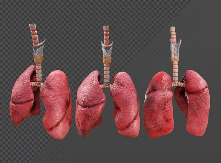
Hemo-pneumothorax is a condition where both blood (hemothorax) and air (pneumothorax) accumulate simultaneously in the distance between the lung and the chest wall, called the pleural space.
Under normal conditions, there is a negative pressure in this cavity, and this pressure allows the lungs to remain attached to the rib cage and remain inflated.
When air or blood enters this cavity from outside, the pressure balance is disrupted, the lung begins to deflate and the fluid/air accumulated in the chest cavity puts pressure on the vessels leading to the heart.
This is an emergency surgical condition that can lead to both respiratory failure and circulatory disorder and must be intervened within minutes.
With modern thoracic surgery protocols, these deposits are quickly evacuated, allowing the lung to swell again and eliminating the risk to life.
What is Hemo-Pneumothorax? How Does It Develop?
Hemo-pneumothorax is a leak into the pleural space as a result of damage to the lung tissue or vessels in the chest wall.
“Pneumothorax” develops when air leaks from the lung, and “hemothorax” develops when blood leaks from the vein; When both are together, the lung is under bilateral pressure.
While the air causes the lung to deflate (collapse); The accumulated blood both compresses the lungs and prepares the ground for shock due to blood loss in the body.
The Relationship Between Hemothorax and Pneumothorax
These two conditions usually trigger each other as a result of a trauma; For example, a broken rib can rupture the lung, leading to both air and blood leakage.
Air leakage rapidly fills the pleural space, while blood settles in the lower parts of this space, pushing the base of the lung upwards.
As a result, the lung becomes dysfunctional, as if being compressed by a vice, and gas exchange reaches a standstill.
Pressure Balance in the Chest Cavity and Lung Collapse
The lungs are elastic like a balloon and tend to deflate as soon as the negative pressure on them is removed.
In hemo-pneumothorax, each breath that enters increases the pressure in the pleural space, making the lung even smaller.
If this condition reaches the stage of “blood pressure pneumothorax”, the heart and main vessels are pushed to the opposite side and blood circulation may stop completely.
What are the Symptoms of Hemo-Pneumothorax?
Symptoms start suddenly and worsen rapidly depending on the severity of the trauma or the amount of blood/air accumulated.
Sudden Severe Chest Pain and Shortness of Breath
Patients often describe a sharp pain as if a knife has been stabbed in their chest, and this pain is exacerbated by breathing.
Since the lung is deflated, the patient begins to feel air hunger; Due to shortness of breath, the patient may have difficulty speaking and may breathe rapidly.
Low Blood Pressure, Heart Rate Acceleration and Shock Symptoms
Due to blood loss (hemothorax), the patient’s color fades and his skin becomes cold and moist.
The body accelerates the pulse to balance the decreasing amount of blood; If the intervention is delayed, blood pressure drops and shock symptoms such as confusion occur.
Causes and Risk Factors of Hemo-Pneumothorax
Although there is usually physical damage behind hemo-pneumothorax, sometimes medical processes can also cause it.
Blunt and Penetrating Chest Traumas
Rib fractures caused by hitting the steering wheel or seat belt pressure in traffic accidents are the most common “blunt” cause.
“Penetrating” traumas such as stabbing or gunshot wounds directly open both the air and blood pathway, leading to a severe picture.
Spontaneous Hemo-Pneumothorax Cases
In rare cases, this condition can develop without trauma as a result of the rupture of a vessel in that area with the bursting of small air sacs (blebs/bullae) in the lung.
It can be seen during menstruation (catamenial), especially in young, thin and tall men or women with endometriosis (chocolate cyst).
Iatrogenic Causes (Conditions After Medical Intervention)
These are accidental injuries that occur during central venous catheter insertion, lung biopsy or fluid removal from the chest cavity (thoracentesis).
Hemo-Pneumothorax Diagnosis and Diagnostic Methods
In emergency room conditions, diagnosis is made within seconds with physical examination and rapid imaging.
Physical Examination: Decreased Breathing Sounds
When the doctor listens with the stethoscope, he hears that the breathing sounds on the affected side have completely disappeared or become very reduced.
In addition, when the rib cage is tapped with a finger (percussion), the drum sound (tympanism) is taken in the upper parts where there is air, and the matite (full sound) is taken in the lower parts where there is blood.
Radiological Imaging: Chest X-ray and Chest CT
In the standing chest X-ray, a flat fluid level (hydroaero line) formed by the blood below is clearly seen.
In stable patients, Computed Tomography (CT) gives the most detailed information to see the source of the bleeding and the extent of lung damage.
Emergency Evaluation with Ultrasonography (FAST)
With the ultrasound method called “E-FAST” in trauma centers, it can be determined whether there is blood or air in the chest cavity within seconds without radiation.
Hemo-Pneumothorax Treatment Methods
The priority of treatment is the “ABC” rule: Open the airway, restore breathing and control circulation (blood loss).
Emergency Intervention: Chest Tube Insertion (Tube Thoracostomy)
The definitive treatment for hemo-pneumothorax is the insertion of a drainage tube through the chest wall into the pleural space.
Thanks to this tube, the trapped air is evacuated and the accumulated blood is taken out; As the pressure is removed, the lung instantly begins to inflate again.
Management of Blood Loss and Fluid Replacement
The amount of blood taken out is monitored and the patient is transfused with fluid and, if necessary, blood through the vein.
If the amount of blood coming from the chest tube is more than 200 ml per hour or exceeds 1500 ml in total, an emergency surgical decision is taken.
Surgical Intervention: When is Thoracotomy or VATS Needed?
Surgery is required in cases where the bleeding does not stop or the tear in the lung does not close with the tube.
Today, these operations can be done by entering through small holes with the closed method (VATS); However, open surgery (thoracotomy) is life-saving in heavy bleeding.
Hemo-Pneumothorax Treatment Decision Chart
| Clinical Status | First Response | The Need for Surgery |
| Small / Stable | Observation + Oxygen | Usually Not Required |
| Intermediate | Chest Tube Insertion | If the leak continues, it is necessary |
| Massive Bleed (>1.5L) | Chest Tube + Blood Transfusion | Emergency Thoracotomy is a Must |
| Recurrent Cases | Chest Tube | VATS (Closed Surgery) Recommended |
Prof. Dr. Levent Alpay: Hemo-pneumothorax is a picture in which the surgeon is racing against time. The most critical mistake here is to just release the air and underestimate the bleeding. As soon as we insert the chest tube, we follow the amount of blood coming in millimeters; Because if there is insidious bleeding inside, this can put the patient in shock. Thanks to VATS technology, which is a closed method, we can now find and repair the bleeding foci of these patients with millimetric cameras without stitches and large incisions. Early intervention prevents the lung from deflating and losing function.
Case Experience (Anonymous):
A 22-year-old young man was brought in with severe pain and bruising on his left side after falling from a height. In the X-ray, it was seen that the left lung was completely deflated and approximately 800 ml of blood accumulated. The lung was inflated with an urgently inserted chest tube, but the air leak did not stop for 3 days. In the VATS (closed surgery) procedure, a burst air sac at the top of the lung was detected and closed, and the patient was sent home with full recovery on the 5th day.
If you have suffered a blow to your chest area or are experiencing sudden shortness of breath, it is vital to consult a specialized thoracic surgeon for immediate evaluation and proper treatment management.
Healing Process and Follow-up After Tube Removal
The chest tube is removed when the air leak is completely stopped and the daily blood supply stops (usually 2-5 days).
After the tube is removed, the patient’s chest X-ray is taken again to confirm that the lung remains swollen.
During the healing process, it is critical for the patient to perform blowing exercises (triflo) to remove any remaining fluids from the lungs and prevent infection.
Frequently Asked Questions
Does Hemo-Pneumothorax Cause Death?
Yes, if the pressure in the pleural cavity reaches a level that puts pressure on the heart (blood pressure picture) or if blood loss cannot be controlled, it can be fatal if not intervened.
Is Pain Felt During Chest Tube Insertion?
The procedure is usually performed under local anesthesia in emergency conditions; Since the area is numbed, no severe pain is felt during the insertion of the tube, but there may be a feeling of pressure.
Is There a Risk of Recurrence After Treatment?
In trauma-related cases, the risk of recurrence after repair of damage is low; However, in cases that develop spontaneously, if there is an underlying structural disorder, there is a risk of recurrence and surgical protection may be required.
Scientific Bibliography
- Cleveland Clinic: Hemopneumothorax Diagnosis and Management
- ScienceDirect: Clinical Overview of Traumatic Hemopneumothorax
- Journal of Trauma and Acute Care Surgery: Chest Tube Management Guidelines
- American Association for the Surgery of Trauma: Thoracic Trauma Protocols