
Ingredients
- What is a Rib Cage Tumor? What are the Types?
- Primary Chest Wall Tumors (Bone and Soft Tissue Origin)
- Metastatic rib cage tumors (spreading from other organs)
- Benign and Malignant Chest Wall Tumors
- Common Benign Masses: Osteochondroma and Lipoma
- Malignant Bone Tumors: Chondrosarcoma and Ewing Sarcoma
- What are the Symptoms of Rib Cage Tumor?
- Swelling and Masses Noticed in the Breast
- Persistent Chest Pain and Shortness of Breath
- Diagnosis and Diagnostic Methods in Rib Cage Tumors
- Imaging with Examination: Thorax CT and PET-CT
- Tru-cut (Needle) Biopsy and Pathological Evaluation
- Rib Cage Tumor Treatment Methods
- Surgical Resection: Complete Tumor Removal
- Chest Wall Reconstruction: Mesh and Titanium Plate Applications
- Postoperative Recovery Process and Oncological Follow-up
- Frequently Asked Questions
- Is Every Swelling in the Rib Cage Cancer?
- Will There Be Deformity After Chest Wall Surgery?
- Is Radiotherapy Required After Tumor Surgery?
- Scientific Bibliography
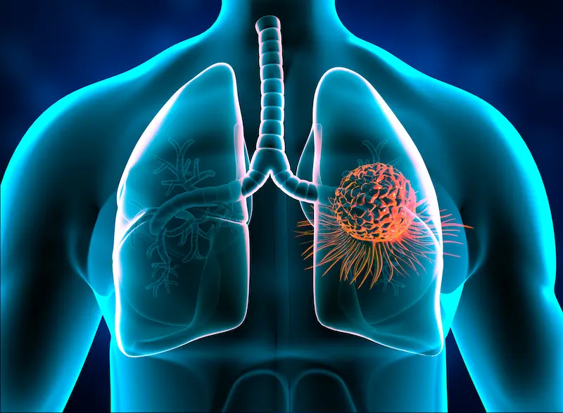
Rib cage tumor, or chest wall tumor with medical name, are masses originating from the ribs, sternum or the soft tissues surrounding them.
Since this region acts as an armor that protects vital organs such as the lungs and heart, tumors that develop here directly affect both structural integrity and respiratory functions.
Although rib cage tumors are rare, their diagnosis and treatment process is quite complex as they can arise from a wide range of different tissue types.
Thanks to modern surgical techniques, most of these tumors can be successfully removed and the chest wall can be reconstructed with artificial materials.
Within the framework of E-E-A-T principles, the most important step in the management of these diseases is to determine the character of the mass at an early stage and to determine the correct surgical margin.
What is a Rib Cage Tumor? What are the Types?
A rib cage tumor is the uncontrolled proliferation of cells in the bone, cartilage, muscle, vessels or nerve tissues that make up the chest wall.
These masses can start directly from the cells of the chest wall itself, or they can occur when cancer from another part of the body spreads there.
When planning treatment, the origin of the tumor and its biological behavior are the most determining factors.
Primary Chest Wall Tumors (Bone and Soft Tissue Origin)
Primary tumors refer to masses that originate directly from the tissues of the rib cage itself.
Approximately 60% of this group is malignant and the rest is benign.
Primary tumors originating from bone tissue are usually located in the ribs, while those originating from soft tissue develop from muscle and adipose tissue.
Metastatic rib cage tumors (spreading from other organs)
Metastatic tumors occur when cancer cells from another organ spread to the chest wall through blood or lymph.
Especially breast cancer, lung cancer, kidney cancer and thyroid cancers are the most common types of metastasis to the rib cage.
Treatment in this case requires a systemic approach that involves not only the mass in the rib cage, but also the main source of cancer.
Benign and Malignant Chest Wall Tumors
Not every mass in the rib cage is cancer; However, a tissue sample and further imaging are essential for precise differentiation.
Benign tumors usually grow slowly and do not show an aggressive tendency to surrounding tissues; But when they increase in size, they can put pressure on the lungs.
Malignant tumors, on the other hand, carry the risk of rapid growth, destruction of bone tissue, and spread to distant organs.
Common Benign Masses: Osteochondroma and Lipoma
The most common benign masses are painless formations that are usually detected incidentally.
- Osteochondroma: They are masses that are a mixture of bone and cartilage, seen at the ends of ribs or long bones.
- Lipoma: They are soft and mobile masses originating from the adipose tissue under the skin; Rarely, they can deepen into the chest cavity.
- Fibrous Dysplasia: It is a condition that weakens the bone, characterized by the disruption of the normal structure of the bone and its replacement by a scar-like tissue.
Malignant Bone Tumors: Chondrosarcoma and Ewing Sarcoma
Malignant tumors constitute one of the most serious intervention areas of thoracic surgery.
- Chondrosarcoma: It is the most common primary malignant chest wall tumor in adults; It originates from cartilage tissue and its complete surgical removal is critical in treatment.
- Ewing Sarcoma: It is more common in children and young adults; It is an aggressive bone cancer that is usually sensitive to chemotherapy and radiotherapy.
- Plasmacytoma: It originates from cells in the bone marrow and often leads to painful destruction of the ribs or sternum.
What are the Symptoms of Rib Cage Tumor?
Symptoms vary depending on the type of tumor, its growth rate and which tissue it affects.
Some tumors do not cause any symptoms for years, while others can cause severe complaints within a few weeks.
Swelling and Masses Noticed in the Breast
The most common reason for application is hard swelling that the patient feels with his hand or can be seen from the outside.
These swellings, which usually start painlessly, may grow over time and cause redness or increased vascularization of the overlying skin.
If the mass is hard and feels attached to the rib, a specialist examination is required immediately.
Persistent Chest Pain and Shortness of Breath
Pain usually occurs as a result of the tumor stretching the bone membrane (periosteum) or pressing on nearby nerves.
- Blunt and Constant Pain: It is more common in malignant masses and can wake you up at night.
- Shortness of Breath: It develops when the tumor grows into the rib cage, preventing the lung from expanding or causing fluid to accumulate in the pleura.
- Cough and Bloody Sputum: It can be seen in advanced cases where the tumor has progressed to the lung tissue.
Prof. Dr. Levent Alpay: Do not neglect every mass in your rib cage by saying “it is a sebaceous gland”. Especially the hard, immobile and recently growing formations that you feel on the ribs should be examined meticulously. In an early diagnosed chest wall tumor, we can not only remove the mass, but also preserve the mechanical structure of the rib cage in the healthiest way. Remember, waiting for the onset of pain may cause you to be late in diagnosis.
Diagnosis and Diagnostic Methods in Rib Cage Tumors
The correct treatment strategy for rib cage tumors begins with determining not only the location of the mass, but also its character and depth relationship with surrounding tissues.
The diagnostic process consists of listening to the patient’s complaints, followed by advanced technology imaging methods and biopsy stages that give definitive results.
A multidisciplinary approach is essential at this stage, as an erroneous evaluation may lead to inadequate surgical margins or unnecessary tissue loss.
Imaging with Examination: Thorax CT and PET-CT
Computed Tomography (CT) is the basic examination that shows the bone destruction in the ribs and sternum, and the distance of the tumor to the lungs in millimeters.
PET-CT, on the other hand, helps us understand whether there is another focus of the tumor throughout the body or the metabolic activity (aggressiveness level) of the mass.
In some soft tissue tumors, contrast-enhanced MRI (MRI) imaging may also be included in the process to see the relationship of the mass with the vascular and nerve packages.
Tru-cut (Needle) Biopsy and Pathological Evaluation
No matter how advanced the imaging methods are, the final step in determining the type of mass is the biopsy.
A small piece of the tumor is taken with a “tru-cut” (thick needle) biopsy, which is usually performed under local anesthesia and under ultrasound or tomography.
This sample is examined in the pathology laboratory to clarify whether the tumor is benign or malignant and the surgical plan is shaped according to this result.
Rib Cage Tumor Treatment Methods
The main therapeutic method in chest wall tumors is the complete removal of the tumor along with some healthy tissue around it.
In order to prevent the tumor from recurrence in malignant masses, a technique called “wide resection” is applied, in which safe surgical margins are preserved.
Surgical Resection: Complete Tumor Removal
During the operation, the ribs or sternum part where the tumor is located are meticulously removed according to the area of spread of the tumor.
The main goal here is to obtain a clean surgical field, leaving no microscopic tumor cells behind.
The size and location of the tumor is the most important factor that determines whether the operation will be performed with the closed (VATS) or open method.
Chest Wall Reconstruction: Mesh and Titanium Plate Applications
The space formed in the rib cage when a large tumor mass is removed must be reconstructed to preserve respiratory mechanics and safety of internal organs.
- Synthetic Meshes: Special patches are used to give flexibility and durability to the chest wall.
- Titanium Plates: Titanium systems replacing the removed ribs maintain the rigidity of the rib cage and allow the lung to swell comfortably.
- Texture Shift (Flep): When necessary, muscle and skin tissues taken from another part of the body are used to cover the repaired area.
Postoperative Recovery Process and Oncological Follow-up
After the surgery, patients undergo a short hospital period during which chest wall stability and breathing capacity are checked.
Thanks to the modern materials used in the repair, patients can gradually return to their daily routines, usually within 1-2 weeks.
In malignant tumors, additional treatments such as radiotherapy or chemotherapy are planned by the oncology council after surgery and the follow-up process is initiated.
Treatment Approaches Comparison Table
| Feature | Benign Tumors | Malignant Tumors |
| Surgical Margin | Removal of the mass only | Wide tissue resection |
| Reconstruction | Usually not required | In most cases, plaque/mesh is required |
| Additional Treatment | Rarely needed | Radiotherapy/Chemotherapy may be required |
| Follow-up Frequency | Annual check-ups | 3-6 months of controls for the first 2 years |
Prof. Dr. Levent Alpay: Rib cage tumor surgeries are not just a mass removal procedure; it is also an engineering work. With titanium plates and special patches that we replace the tissues we have removed, we restore the natural resistance and appearance of your rib cage as if you had never had surgery. Our most important assistant in this process is error-free surgical planning with advanced imaging techniques.
Case Experience (Anonymous):
In the examinations performed on our 50-year-old patient, who noticed a painful swelling in his right rib, a 6 cm chondrosarcoma (malignant mass) originating from the rib was detected. The tumor was removed within wide limits along with the three ribs it affected. The resulting cavity was reconstructed with titanium plates and a special patch. Our patient, who did not experience respiratory distress after the surgery, continues to be followed up in his 5th year in a completely healthy and tumor-free manner.
For a suspicious mass or persistent pain you notice in your chest area, you can make an appointment with our clinic and seek expert opinion for the correct diagnosis and personalized treatment planning.
Frequently Asked Questions
Is Every Swelling in the Rib Cage Cancer?
No, a significant portion of swelling in the rib cage is lipoma (sebaceous gland), cyst, or benign bone growths; However, for definitive differentiation, specialist examination and imaging are absolutely necessary.
Will There Be Deformity After Chest Wall Surgery?
Thanks to modern reconstruction techniques (titanium plates and mesh), the area where the mass is removed is repaired in accordance with its former form and a significant deformity is prevented.
Is Radiotherapy Required After Tumor Surgery?
Depending on the type of tumor, the condition and stage of the surgical margins, additional radiotherapy may be recommended by oncology specialists to reduce the risk of recurrence of the disease.
Scientific Bibliography
- The Annals of Thoracic Surgery: Chest Wall Tumors: Diagnosis and Management
- PubMed (NCBI): Primary Malignant Chest Wall Tumors
- Journal of Thoracic Disease: Reconstruction Techniques in Chest Wall Surgery
- Lancet Oncology: Systemic Therapy and Surgical Approaches for Sarcomas