
Ingredients
- What is the Pleura?
- What is Fluid Collection in the Pleural Membrane?
- What are the Causes of Fluid Collection in the Pleura?
- What are the Symptoms of Fluid Collection in the Pleura?
- Is Fluid Collection in the Pleura Dangerous?
- Pleural Cancer (Malignant Pleural Mesothelioma)
- Pleural Inflammation (Empyema)
- Thickening of the Pleura and Its Causes
- Diagnostic Methods in Pleural Diseases
- Pleural Biopsy and Thoracentesis
- Pleural Surgeries and Treatment Methods
- Closed Method Pleural Surgery (VATS – Thoracoscopic Surgery)
- Pleurodesis (Pleural Bonding) Procedure
- Decortication Surgery
- Frequently Asked Questions
- How long does pleural surgery take?
- What happens if the pleura is removed?
- Why Does Fluid Accumulation in the Lungs Recur?
Lung and pleural surgery is a surgical specialty that aims to correct structural or functional disorders of the lung tissue in the rib cage and the protective membrane surrounding this tissue (pleura).
This surgical branch aims to improve the quality of life and preserve respiratory functions by using all the possibilities of modern medicine both in the treatment of benign diseases and in the management of aggressive processes such as cancer.
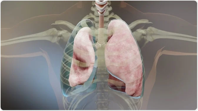
What is the Pleura?
The pleura is a serous membrane consisting of two parallel layers that surrounds the lungs and covers the inner surface of the chest wall.
Between these two layers of membranes is a very small amount (about 15-20 ml) of lubricating fluid that allows the lungs to slide without friction in the rib cage during breathing.
The pleura is not just a covering but a critical barrier that maintains the integrity of the lung and maintains intrathoracic pressure balance.
What is Fluid Collection in the Pleural Membrane?
Pleural effusion is a condition in which a much larger amount of fluid accumulates in the space between the two pleuras.
This condition, popularly known as “blistering in the lung”, can prevent the lung from fully swelling, leading to severe respiratory distress.
Fluid buildup is not considered a disease in itself, but often a symptom of another systemic or local pathology in the body.
What are the Causes of Fluid Collection in the Pleura?
The accumulation of fluid in the pleura can be triggered by many different mechanisms:
- Heart and Liver Problems: Conditions like heart failure or cirrhosis can disrupt the balance of pressure in the body, leading to fluid leakage.
- Infections: Fluids that develop after pneumonia are among the most common causes.
- Cancers: Tumors like lung cancer or mesothelioma can increase fluid production directly on the membrane.
- Renal Impairment: As a result of the body’s inability to regulate fluid balance, an increase in fluid may occur in the pleural space.
- Pulmonary Embolism: Blockages in the pulmonary vessels can lead to effusion.
What are the Symptoms of Fluid Collection in the Pleura?
Depending on the amount and speed of the accumulated fluid, the symptoms may be exacerbated:
- Shortness of Breath: It is the most prominent symptom that develops due to decreased lung capacity.
- Chest Pain: Stinging pains felt especially when breathing deeply are typical.
- Cough: It is often accompanied by a dry cough without phlegm.
- Fever and Sweating: If the fluid is due to an infection (empyema), high fever is seen.
According to Prof. Dr. Levent Alpay; Patients often express difficulty breathing while lying on their backs; If you have unilateral pain and feel like a stabbing sensation when breathing deeply, this may be a sign of pleural irritation.
Is Fluid Collection in the Pleura Dangerous?
Fluid collection in the lungs can be life-threatening depending on the underlying cause.
While small amounts of fluids can be monitored, rapidly accumulating or inflamed fluids can cause lung deflation and severe pictures such as sepsis.
Especially in cases where cancer is suspected, the character of the fluid is critical as it determines the stage of the disease.
Pleural Cancer (Malignant Pleural Mesothelioma)
Malignant Pleural Mesothelioma is an aggressive type of cancer that originates from the lung membrane’s own tissue, usually developing as a result of exposure to asbestos fibers.
In this disease, the pleura becomes thickened, irregular and wraps around the lung like armor, making breathing almost impossible.
Early diagnosis and multidisciplinary surgical approach are the most basic elements to increase survival time.
Pleural Inflammation (Empyema)
Empyema is a buildup of inflamed fluid or pus trapped in the pleural space.
It usually develops after untreated or inadequately treated cases of pneumonia.
If this inflamed tissue is not evacuated in time, it leads to permanent thickening of the pleura and trapping of the lung.
Thickening of the Pleura and Its Causes
Pleural thickening is a condition in which the membrane loses its elasticity and hardens.
The most common causes include past tuberculosis (tuberculosis), empyema, asbestos exposure or incomplete cleaning of blood leaking into the pleural cavity (hemothorax).
The thickened membrane restricts the expansion of the lung, causing chronic shortness of breath and narrowing of the rib cage.
Diagnostic Methods in Pleural Diseases
In the modern diagnostic process, imaging and interventional methods are used together:
- Chest X-ray and Tomography (CT): It is the first step to see the amount of fluid and the structure of the membrane.
- Ultrasonography (USG): It is a safe guide to determine the exact location of the fluid and intervene with the needle.
- PET-CT: It is preferred to measure metabolic activity in cases where cancer is suspected.
Pleural Biopsy and Thoracentesis
Thoracentesis: It is the process of taking or draining the fluid for sample purposes by entering a special needle between the lung membranes.
Pleural Biopsy: In cases where fluid examination is insufficient, a small piece of pleural tissue is taken and sent for pathological examination.
These procedures can usually be performed under local anesthesia without requiring hospitalization.
Pleural Surgeries and Treatment Methods
The main goal in the treatment of pleural diseases is to re-release the lung and stop fluid accumulation.
| Method | Purpose | Method of administration |
| VATS (Off) | Diagnosis and treatment | Camera accompaniment through small holes |
| Pleurodesis | Preventing fluid recurrence | Gluing two membranes together |
| Decortication | Releasing the lung | Peeling off the thickened membrane |
Closed Method Pleural Surgery (VATS – Thoracoscopic Surgery)
VATS is a modern surgical method performed with the help of a camera and special tools through 1 to 3 small holes without making large incisions in the rib cage.
It is the gold standard today because it has less pain, shorter hospital stay and better aesthetic results.
According to Prof. Dr. Levent Alpay; Closed surgery is not only comfortable, but also significantly accelerates the patient’s adaptation to normal life after treatment, thanks to the fact that the immune system is less affected.
Pleurodesis (Pleural Bonding) Procedure
It is the process of closing this gap by injecting a sterile substance (for example, talcum powder) between two pleural leaves, especially in patients who constantly collect fluid due to cancer.
The aim is to leave no space for fluid accumulation and to save the patient from the trouble of constantly coming to the hospital and draining fluids.
Decortication Surgery
In cases where the pleura is excessively thickened and squeezes the lung like a cage, this is the process of peeling off the hardened layer from the lung tissue with millimeter meticulousness.
After this surgery, the lung can expand again and a significant increase in the patient’s breathing capacity is achieved.
Clinical Experience and Case Example (Anonymous):
A 45-year-old male patient who developed severe fluid accumulation and membrane thickening in the rib cage after persistent pneumonia did not respond to drug treatment. In the patient who was entered with VATS (closed method), the inflamed tissues trapping the lung were cleaned by performing the “Decortication” procedure, and the patient was discharged with full respiratory capacity 4 days after the operation.
Frequently Asked Questions
How long does pleural surgery take?
Although it varies depending on the scope of the procedure, closed (VATS) methods are usually completed between 45 and 90 minutes; More extensive surgeries such as decortication may take 2-3 hours.
What happens if the pleura is removed?
Removal of part or all of the pleura (pleurectomy) does not cause loss of life; Over time, the body harmonizes this space with the chest wall tissue, and respiratory function usually improves.
Why Does Fluid Accumulation in the Lungs Recur?
If the main disease causing fluid collection (cancer, heart failure or chronic infection) is not controlled or if bonding (pleurodesis) is not performed between the membranes, fluid may accumulate again.
Scientific Bibliography
- The Lancet: Pleural effusion: a clinical review – Comprehensive analysis on the clinical management of pleural fluids.
- PubMed (NCBI): Management of Malignant Pleural Effusions – Surgical and medical treatment methods of malignant pleural effusions.
- Journal of Thoracic Disease: Video-assisted thoracoscopic surgery (VATS) for pleural diseases – The efficacy and results of closed surgical methods in pleural diseases.
- European Respiratory Review: Pleural infection: past, present and future – Historical development of empyema and pleural infections and modern treatment approaches.